Walking on the side of researchers striving to establish an assay for the prediction of  the response of human lung cancer patients to cancer immunotherapy, we have established a 3D microfluidic tumour microculture system that involves the seeding of LLC (Lewis Lung Cancer) syngeneic mouse tumour aggregates in the central hydrogel channel of an Aim Microfluidic Chip  and the injection of anti-PD1 or anti-PDL1 antibodies in the two  flanking side channels.

Proteomics analysis  proteomics of the untreated vs anti-PD1 treated tumour samples revealed the signature of the response to treatment (A). Metascape analysis of increased  (B) or decreased ( C) protein expression after anti-PD1 treatment revealed the biological pathways involved.

With this data we have the proof of concept to support that a similar humanized PDL1/PD1 system can be used for screening the responses of human patients to anti-human PD1 therapeutics.

This work was performed under the European funded program “Next Generation EU” Greece 2.0, https://greece20.gov.gr/en/) BioOnChip, “Development of a Bronchoscopic Biopsies-On-Chip platform for immunotherapy drug screening in non-small cell lung cancer” that aimed to explore the applicability of an innovative 3D microfluidic microculture in predicting real-time responses to PD1-blockade in NSCLC patients, that, if efficient, might have important socioeconomic impact.

Read More

We are excited to introduce a new addition to our collection of genetically modified mice used in human disease modelling. We have now developed and characterized a proprietary Rag1KO/BMC mouse line that either alone or in combination with our collection of proprietary mouse lines allows us to offer a new series of anti-CD40 colitis mouse models that can serve as invaluable preclinical tools to better understand and treat the pathogenic mechanisms involved in the development and progression of Inflammatory Bowel Disease (IBD).

💉 The anti-CD40 model of colitis is a valuable model to study innate immune responses in colon inflammation and to evaluate the therapeutic effect of TNF, IFNγ or IL-12/IL-23 p40 inhibition 💊

What is the anti-CD40 induced colitis model?
Activation of CD40 by an agonistic anti-CD40 antibody in mice lacking T and B cells, as the Rag1 or Rag2 knockout mice, leads to the activation of innate immune responses, the excessive production of IL-23, IL-1β and IL-12 and the development of inflammation in the colon.
The anti-CD40 induced colitis pathology is characterized by body weight loss, increased levels of circulating cytokines, reduced colon length and increased colon weight,  accompanied by relevant histopathological signs of gut inflammation and extensive crypt destruction.


Read More

Biomedcode is in search  of a motivated Research and Business Manager to join the team and lead strategic initiatives focused on drug discovery projects. S/he will focus on identifying and pursuing grant funding and investment opportunities, managing intellectual property (IP) portfolios and liaising with potential stakeholders. The ideal candidate will lead initiatives to generate and engage with business partners to exploit the company’s IP portfolio. Critical thinking and a demonstrated ability to execute a particular IP exploitation strategy, identify partnerships and financing opportunities in the life sciences industry  as well as develop and manage IP commercialization strategies are required.

Read More

This newest addition in our collection of humanized mouse disease models integrates characteristics of the human Systemic Lupus Erythematosus complexity and develops a chronic multiorgan autoimmune disease marked by proteinuria, anti-dsDNA antibodies, severe inflammatory lesions in the skin and milder pathologies in the kidneys and lungs.

This novel model of lupus can prove to be an invaluable translational tool for studying the aetiopathogenic role of the IL23 cytokine in SLE and for use as a preclinical tool to assess the efficacy of novel  lupus  therapeutics.

Published in Arthritis Rheumatol. 2024 Feb 15. doi: 10.1002/art.42830.

Read More

Idiopathic pulmonary fibrosis (IPF) is a chronic and progressive interstitial lung disease characterized by irreversible fibrosis. It involves the transition of fibroblasts to myofibroblasts, the tissue stiffening and alveolar epithelium injury eventualy causing irreversible and progressive lung damage.

Various methods have been employed to simulate pulmonary fibrosis for the purpose of evaluating potential anti-fibrotic therapeutic treatments. Apart from the use of IPF animal models where the pathology is induced through the endotracheal administration of bleomycin and the development of 2D cell cultures for the study of fibrosis mechanisms, three-dimensional (3D) cultures have also emerged as valuable tools for modeling IPF.

One such 3D culture approach is the generation of precision-cut lung slices (PCLS), where thin slices with precise thickness are obtained from fresh tissue with the use of a vibrating microtome. These slices recapitulate the in vivo lung environment by offering a preserved tissue structure with intact cell-cell and cell-extracellular matrix interactions, allowing for the study of molecular mechanisms between different lung cell populations and facilitating lung degeneration and alveologenesis. The advantage of this approach is that it allows for the generation of multiple slices from the lungs of a single mouse , thereby greatly reducing the number of animals required for individual experiments.

As it has been shown that PCLS obtained from IPF patients or bleomycin-induced mouse lungs, exhibit reduced expression of fibrotic markers following TGF-β inhibition, Biomedcode is currently developing a preclinical ex vivo tool using PCLS derived from bleomycin-induced mouse lungs to screen ex vivo the therapeutic efficacy of anti-fibrotic drugs. Using Nintedanib, an intracellular tyrosine kinase inhibitor that targets fibroblast recruitment, proliferation and differentiation, as a positive control and through the assessment of fibrosis biomarkers such as fibronectin, alpha Sma and collagen 1A1, we have generated experimental evidence supporting that PCLS from IPF-affected mouse lungs can nicely serve as a powerful preclinical tool to assess the anti-fibrotic effect of test articles targeting inflammation and fibrosis.


Graph created by bioRender

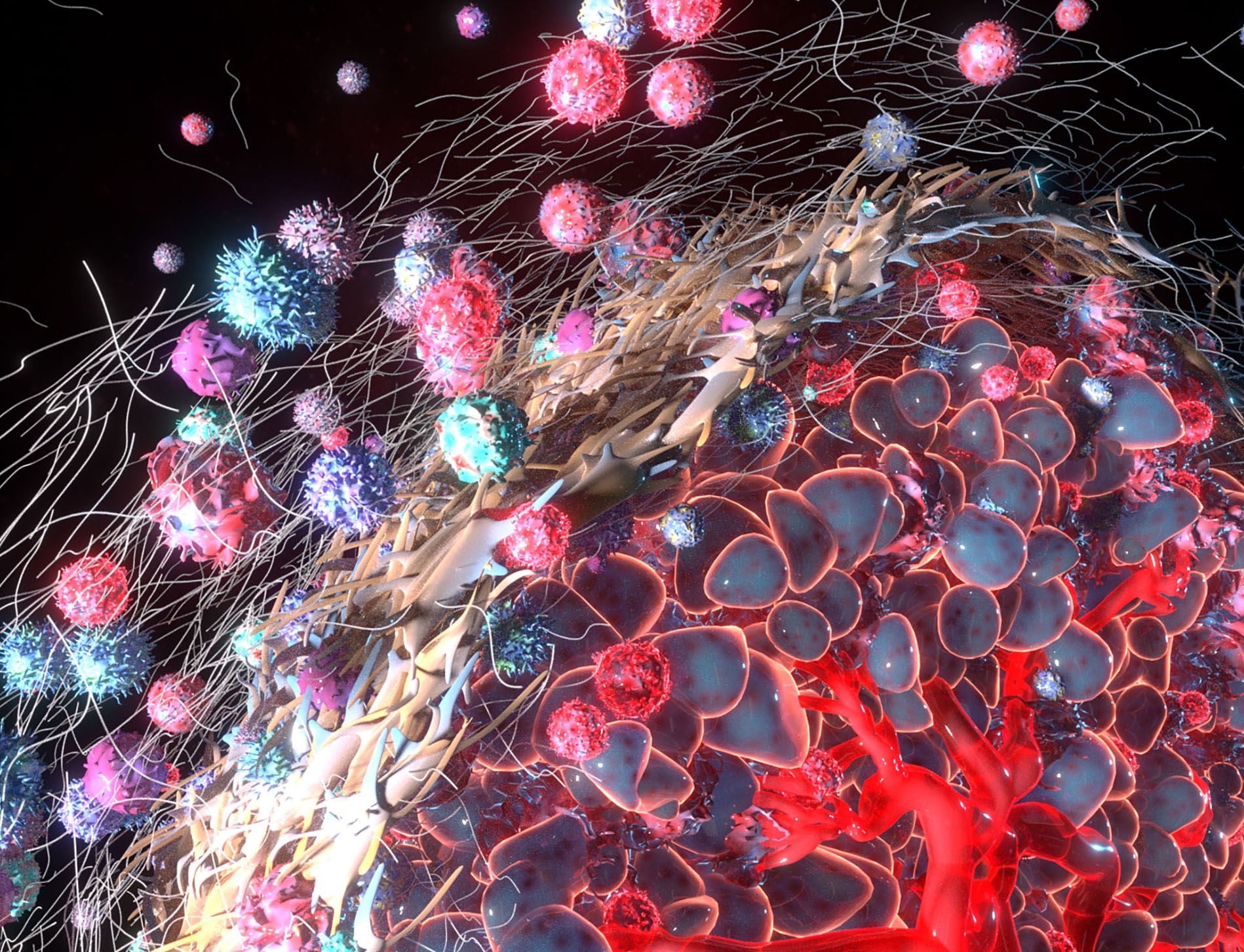
TNF is a pleiotropic cytokine serving important functions in pathophysiology, as it plays regulatory role in the growth, differentiation and death of immune and non-immune cells. Its functions are mediated through binding to two transmembrane receptors, TNFR1 and TNFR2, that deliver signals to activate a variety of responses ranging from proliferation to apoptosis and necrosis.

Dysregulation of TNF has been implicated in immune mediated inflammatory diseases including rheumatoid arthritis, inflammatory bowel disease, psoriasis, scleroderma, systemic lupus erythematosus, atherosclerosis as well as autoimmune pathologies, cancer etc.

Since the approval of the first TNF inhibitors, back in 1998, TNF blockade has been used as a successful approach in treating a broad range of pathologies, having nonetheless the expected compromises stemming from the severe side effects that result from the blockade of the whole range of the physiological functions of TNF, with the increased susceptibility to infections being one of the majordrawbacks of this therapeutic approach.

The pioneering work of two Greek scientists, Kassiotis and Kollias, published in 2001 in JEM (193:427) and JCI (107:1507), shed light for the first time on the importance ofuncoupling the TNFR1- and TNFR2-TNF signaling pathways. This work  proposed that specific blockade of TNFR1 signaling could offer advantageous novel therapeutic approaches as they would suppress the TNF pro-inflammatory activity, leaving unaffected its TNFR2-dependent immunosuppressive activity. In the following years, indeed TNFR1 emerged as an attractive target towards ameliorating the detrimental effects of the TNF signalling pathway and various TNFR1 inhibitors are currently under development or in clinical trials.

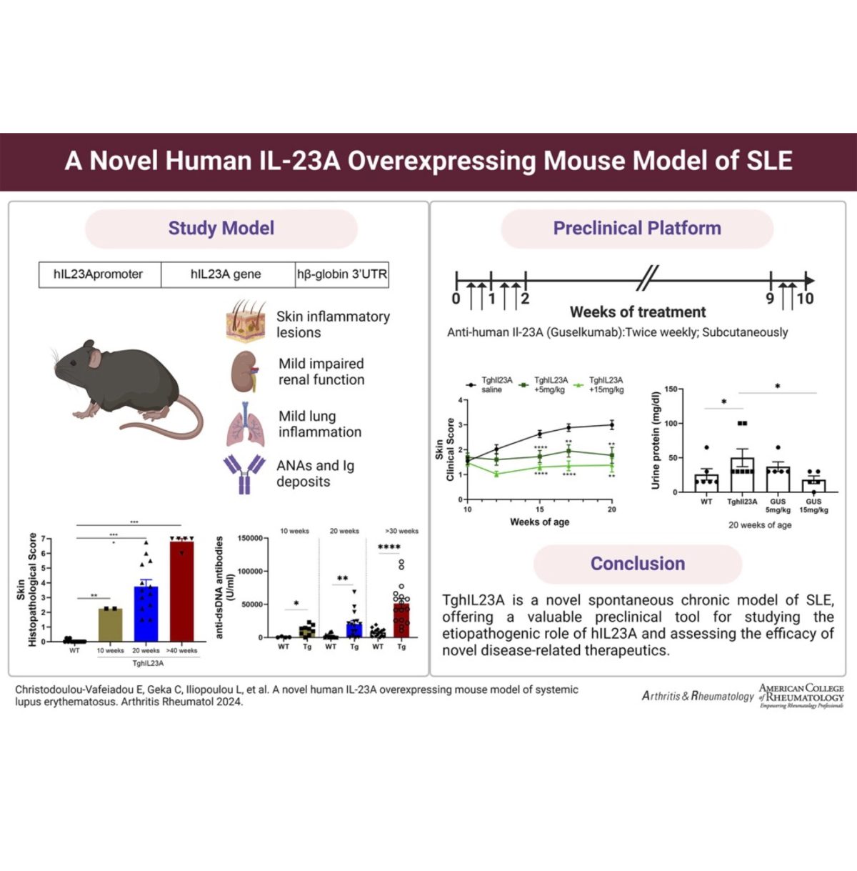
A unique tool for testing anti-hTNFR1 therapeutics is the human TNFR1 knock in (hTNFR1KI) mouse model, that expresses human TNFR1 in the absence of its mouse counterpart. The hTNFR1KI mouse combined, as shown in the table below, with induced or spontaneous disease models offers a fully equipped armory for the evaluation of human therapeutics targeting TNFR1.

Preclinical Platforms for the evaluation therapeutics targeting human TNFR1

Model Duration Read outs
Experimental Autoimmune Encephalomyelitis

MOG-induced EAE in hTNFR1KI mice

25-30 days -Weight

-Clinical Scoring

-Histology

Spontaneous Arthritis 

Tg197hTNFR1KI

TNFΔARE/+ hTNFR1KI

12 weeks

4-8 weeks

-Weight

-Clinical Scoring

-Histology

Induced Arthritis 

Collagen Antibody Induced Arthritis (CAIA) in hTNFR1KI mice

2 weeks -Weight

-Clinical Scoring

-Histology

-Serum Cytokine levels

Acute Inflammation

TNF/GaIN induced inflammation in hTNFR1KI mice

24 hours -Serum cytokine levels-Survival

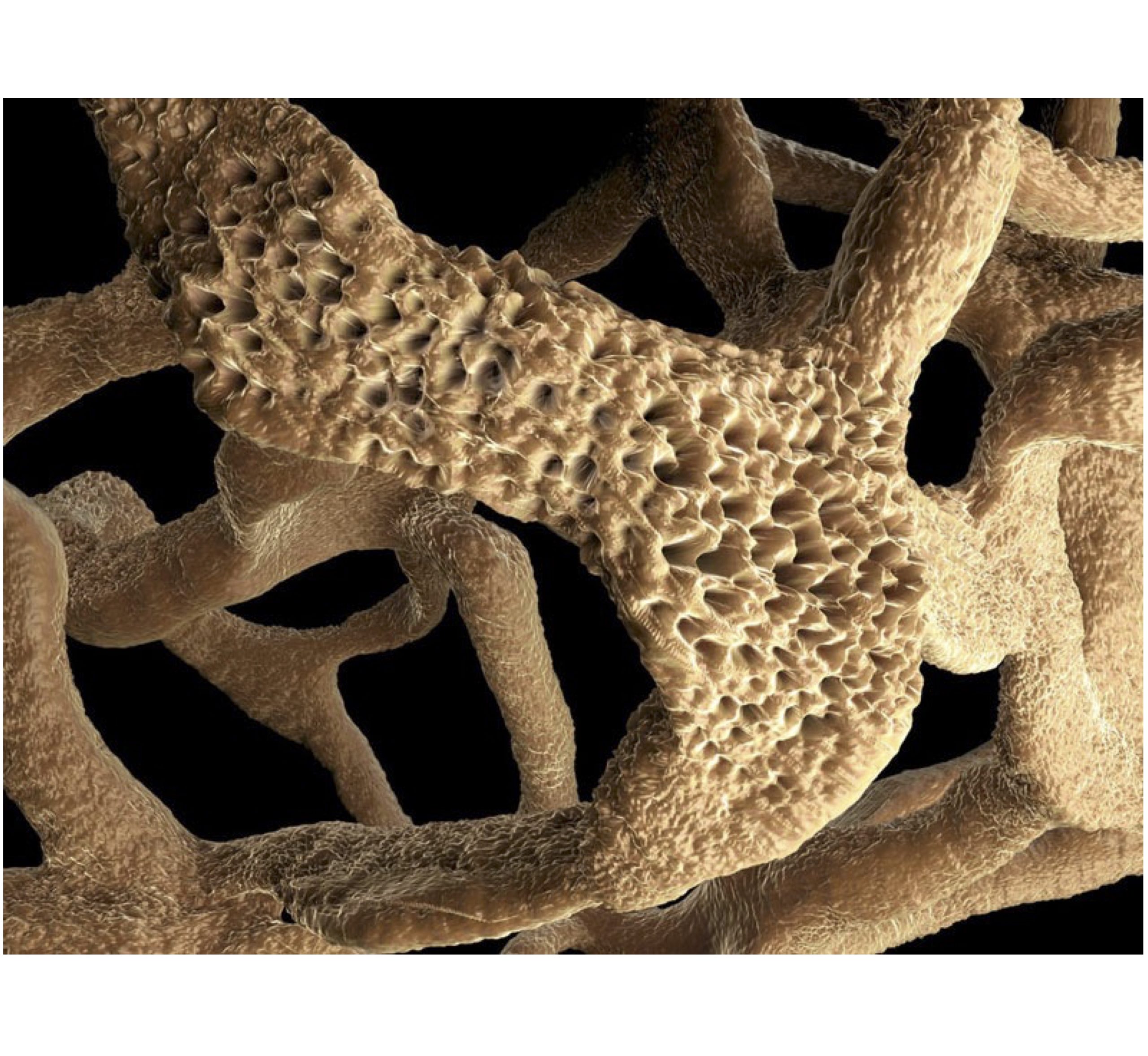
One of the models described above that has a particular interest due to its complexity and similarity to the human condition is the TNFΔAREhTNFR1KI mouse model that due to the dysregulated expression of TNF develops spontaneously intestinal inflammation, arthritis and cardiovascular disease, perfectly recapitulating many of the features that constitute the complexity of the human disease. TNFΔARE was the first mouse model that unequivocally established the causal role of TNF in the inflammatory response of Crohn’s-like ileitis, while further studies revealed numerous promising therapeutic targets some of which were later developed into approved drugs, such as Ustekinumab, vedolizumab, and natalizumab.

The TNFΔAREhTNFR1KI mouse model, faithfully reproduces all TNFΔARE phenotypes and is an ideal tool for translational research, supporting the drug discovery and development of biologics and small molecules targeting TNF, hTNFR1 as well as other potential therapeutic targets of Crohn’s-like ileitis.

Fibroblasts are responsible for creating and maintaining the extracellular matrix that normally supports all the connective tissues. Tumors have the ability to recruit fibroblasts (Cancer Associated Fibroblasts (CAFs)) that develop a fibrotic microenvironment that supports their growth while protecting them from the immune system. This fibroblast-generated scar tissue is associated with the reduced efficiency of chemotherapies and poor outcome of treatment.

A new publication in Nature Cancer provides evidence that reducing scar tissue with the PXS-5505 small molecule inhibitor can help to treat pancreatic cancer by making it more accessible to chemotherapy treatment. PXS-5505 is an inhibitor of the lysyl oxidase family of enzymes which causes collagen to build up around tumors creating fibrotic scar tissue that acts as a barrier inhibiting the access of chemotherapy agents. The study offers exciting evidence that therapeutic strategies that involve the targeting of tumor-associated stroma can offer several benefits to the treatment of cancer including improved chemotherapy responses, reduced metastatic burden and more importantly prolonged survival.

Read the full article in Nature Cancer